

Visualization
Grid
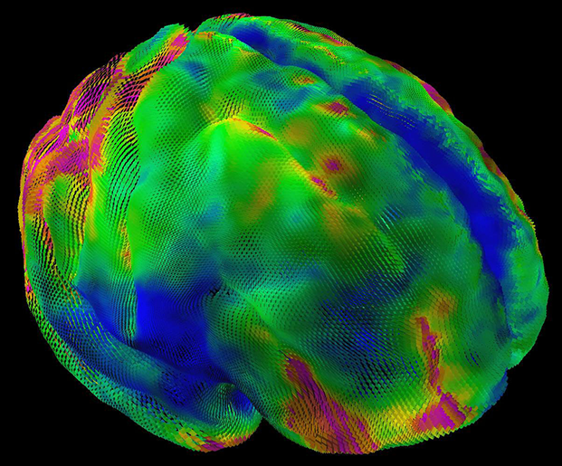
Diffusion Tensor Imaging of a Healthy Brain
Diffusion Tensor Imaging (DTI) shows the direction of water molecules along brain neurons, the direction of which are differentiated by color.
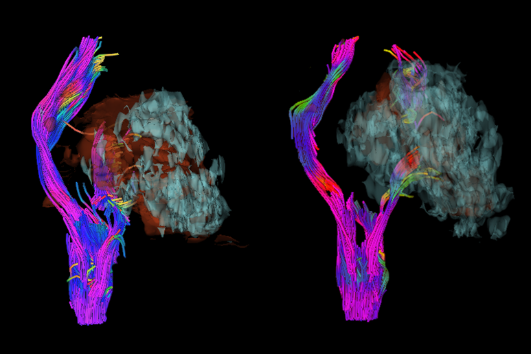
Comparison of Acute and Chronic Brain Hemorrage
The left image shows the brain shortly after injury, the right after treatment. Comparison of the images shows the damage to neurons as a result of the injury.
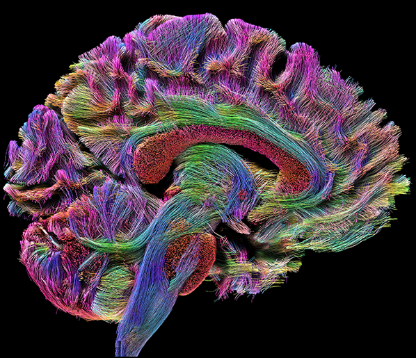
Tractography - Sagittal
A sagittal view of diffusion tractography of a single subject.
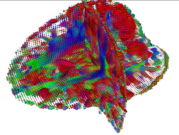
Diffusion Tensor Imaging of a Healthy Brain
Diffusion Tensor Imaging (DTI) shows the direction of water molecules along brain neurons, the direction of which are differentiated by color.
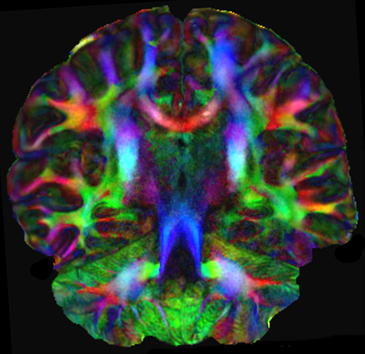
Diffusion Tensor Imaging of a Healthy Brain
Diffusion Tensor Imaging (DTI) shows the direction of water molecules along brain neurons, the direction of which are differentiated by color.
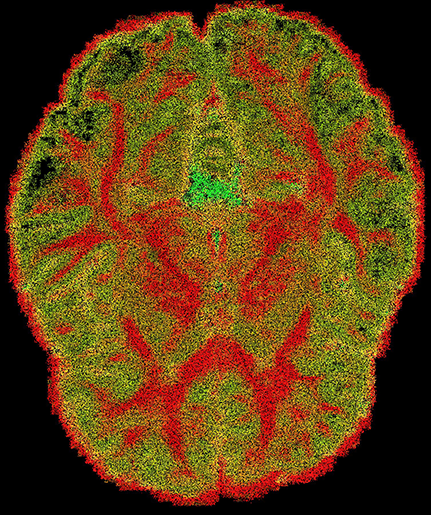
Diffusion Anisotropy
Diffusion Ansiotropy shown by color, red is higher ansiotropy.
Tractography - Axial
An axial view of diffusion tractography of a single subject.
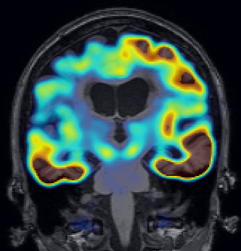
PET Scan of Alzheimer's Brain
A Positron Emission Tomography (PET) scan of a brain with Alzheimer's Disease.
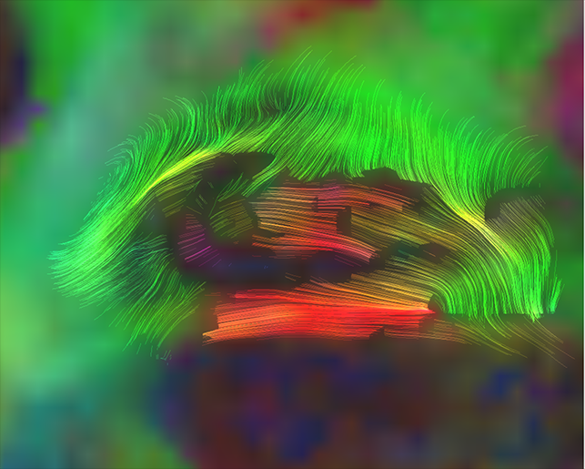
Fibers of the Hippocampus
Tractography fibers of the hippocampus in a healthy brain.
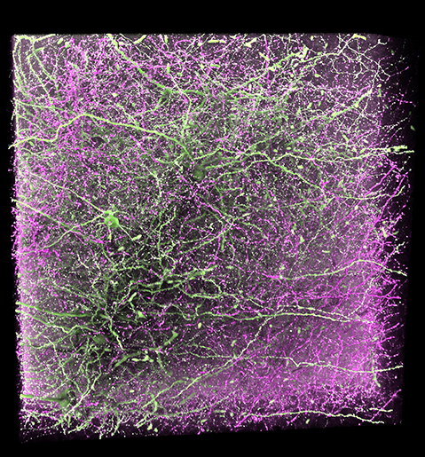
Prefrontal Circutry
Microscopic data of neuronal connections of the medial prefrontal cortex to (green) and from (purple) the amygdala.
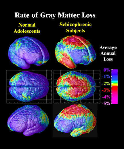
Comparison of Healthy and Schizophrenic Brains
A comparison of the grey matter in the brains of adolecents with and without schizophrenia.
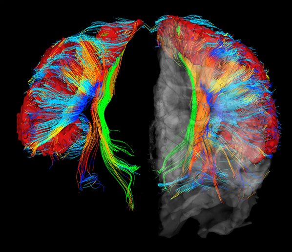
White Matter of S1 - Frontal
The green fiber tracts shown are the neurons of the primary somatosensory cortex (S1) which are functionally involved in processing signals from the GI tract.
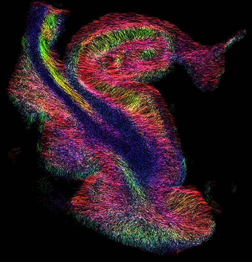
Alzheimer's Hippocampus Tractography
Tractography of the hippocampus in a brain with Alzheimer's disease.
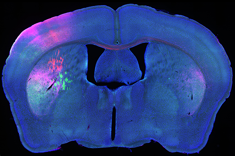
Mouse Mid-brain Fluorescent Tract Tracing - Coronal
A coronal slice of the mid-brain of a mouse. Florescent dye is used to show how neural tracts are connected across hemispheres.
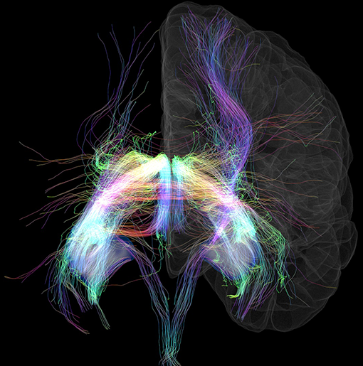
Hippocampal Connectivity
Diffusion tracts showing connections with the hippocampus.
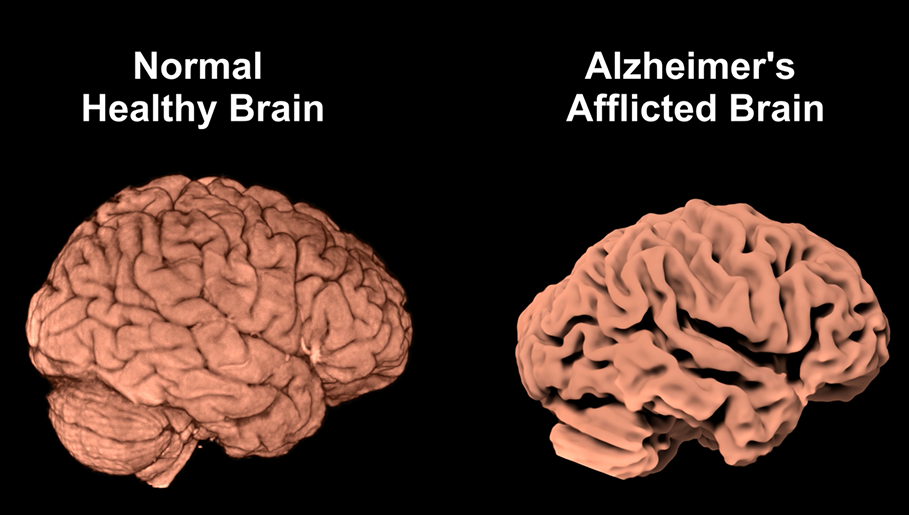
Comparison of Normal and Alzheimer's Brains
Rendering of the brain from imaging data showing the loss of white matter in Alzheimer's Disease.
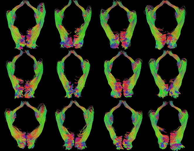
Tractography of Visual Pathways from Multiple Subjects
Inferior?posterior views of the tractography of 12 visual pathways reconstructed from 12 different subjects.
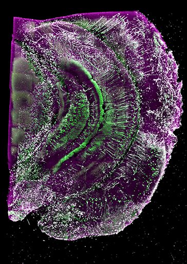
Hippocampal Mouse Neurons
A 3D rendering of microscopic data collected from the mouse hippocampus.
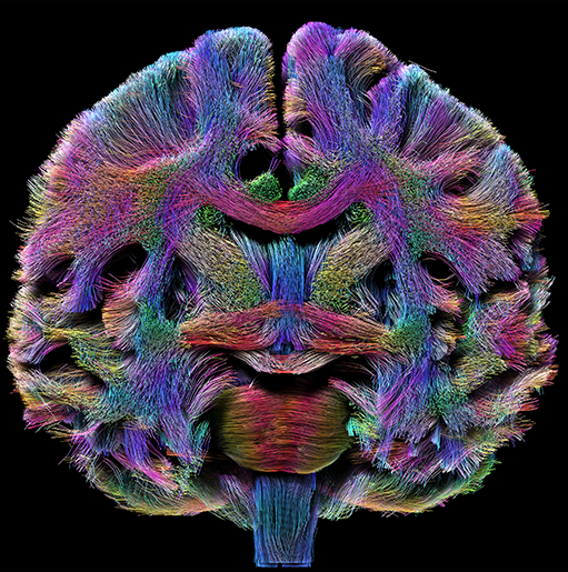
Tractography - Coronal
A coronal view of diffusion tractography of a single subject.
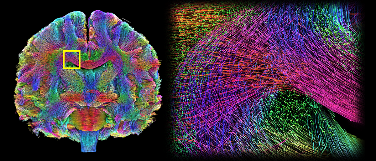
Multiscale Tractography
A multiscale viewpoint of diffusion tractography.
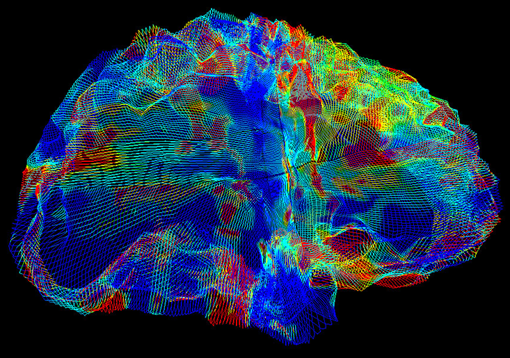
Diffusion Tensor Imaging of a Brain with Alzheimer's
Diffusion Tensor Imaging (DTI) shows the direction of water molecules along brain neurons, the direction of which are differentiated by color.
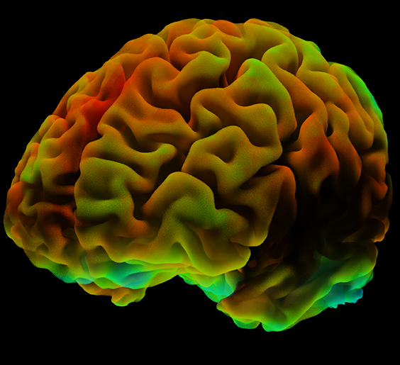
fMRI Brain Activity
A snapshot of fMRI brain activity.