
LONI Brain Parser
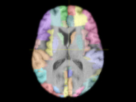
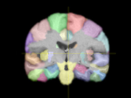
Brain Parser software segments regions of interest based on a training set of data and generates 3D MRI volumes. The software comes pre-trained on a provided data set but can be retrained to work with your desired regions of interest.
Visit Forum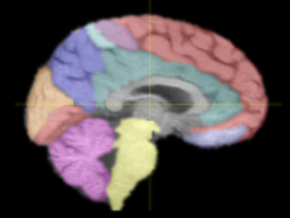
Features
- Awarded the "Best Automated Segmentation" Award at the 2007 International Medical Image Computing and Computer Assisted Intervention conference
- Learning based and highly adaptive
- Efficient computation, whole brain segmentation in 30 minutes or less
- Seamless integration of image appearance and context
Description
Brain Parser software segments regions of interest based on a training set of data and generates 3D MRI volumes. The software comes pre-trained on a provided data set but can be retained to work with your desired regions of interest. Brain Parser specifically includes skull and scalp removal, image nonuniformity compensation, voxel-based tissue classification, topological correction, rendering, and editing functions. The collection of tools is designed to require minimal user interaction to produce cortical representations. Surface representations of the human cerebral cortex are important for visualization and analysis of neuroimaging data (Thompson et al., 1997) and as constraints for localizing functional activation from magneto-encephalography (MEG) and electro-encephalography (EEG) data (Dale and Baillet). MR imaging offers neuroanatomical detail, but extraction of cortical surfaces from MR imagery faces several problems, including measurement noise, partial volume effects, and image nonuniformity do to magnetic field inhomogeneities.Usage
The following steps are required to go from raw data to fully segmented data: <ul> <li>Data must be in Analyze Image 16 bit short format and should have a reasonable intensity range from 0 to 5000</li> <li>Data must be aligned to the template image</li> <li>Skull stripping</li> <li>Registration</li> <li>Intensity normalization</li> <li>After you run LONI Brain Parser you will need to invert your registration to match the orientation of the original data</li> </ul> If you have good quality data that approximates the template image, your results are likely to be more successful.<br /> <br /> Alternatively, you can run the LONI Brain Parser-Pipeline Workflow in our <a target=\"_blank\" href=\"demo.php?id=69\">Try it now</a> area. The workflow has been setup with the pre-processing and post-processing steps predefined for ease of use.
System Requirements
Windows
- Version: 2.0
- Size: 4 MB
- OS: Windows 32 bit
- Processor: 2.8MHz processor or higher
- Memory: 2 GB
Linux
- Version: 2.0
- Size: 4 MB
- OS: Linux 32 bit
- Processor: 2.8MHz processor or higher
- Memory: 2 GB
Installation
WindowsDownload the Windows exe and double click to launch.
Linux
Download the Linux binary and double click to launch.
Via the LONI Pipeline Workflow
The Try it now area offers a test drive of the LONI Pipeline-Brain Parser Workflow. You can try out your data with all the processing steps set up for you. This will require Java 1.5 or higher. If you have the LONI Pipeline running locally on your machine you can download the .pipe file. You can hit "Play" which will connect you to our guest computer cluster. Otherwise, you can edit the "Connection" details and login to the main cluster if you have already gained access. If you would like to use our cluster but do not currently have access, you may request it on the LONI Pipeline website.
Purpose
LONI Brain Parser allows the user to quickly segment anatomical brain structures using automated delineation methods. The software can also be "trained" to locate areas of interest in the brain that it is not programmed to locate by default. Whereas segmentation by hand can take hours or even days, auto-segmentation only takes about half an hour. With this segmented data researchers can study the shape and morphometry of the structures to discover new patterns and associations in areas such as development and disease.